脑积水Hydrocephalus · 脑 · 神经系统 · NTP ntp-0393
诊断要点:在 NTP 研究中,应尽可能确定脑积水的病因。除非脑积水继发于肿瘤阻塞,否则应诊断为脑积水。
病变描述
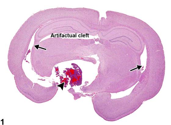
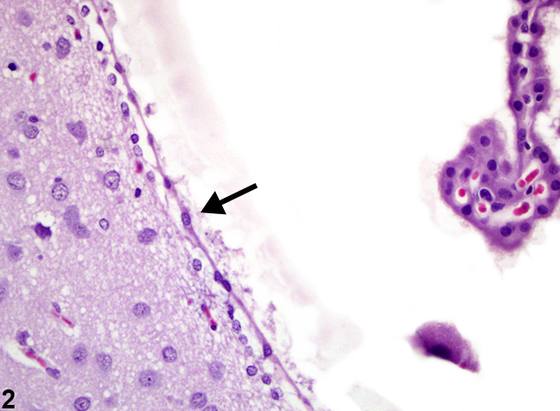
脑积水(hydrocephalus)可为交通性或非交通性;也就是说,前者没有明显的梗阻过程,而后者在脑室连接通路的某处存在梗阻性病因。最常见的是,交通性脑积水被认为由脑脊液生成的特发性增加或吸收减少所致。然而,大多数病例并未得到明确的探究。Figure 1 是一例非交通性、梗阻性脑积水,侧脑室(箭头)因垂体肿瘤(arrowhead)阻塞第三脑室腹侧而继发扩张。Figure 2 描绘了正常立方状室管膜细胞(ependymal cell)(箭头)变扁的衰减改变。这是由于与偶然发现的垂体囊肿相关的梗阻性脑积水性室室扩张所致。在此例中,脑积水时脑室内压增高对室管膜细胞的影响显而易见。正常的立方状室管膜细胞被拉伸以覆盖增加的脑室表面积。上皮与下方脑组织之间存在一定程度的水肿性分离,邻近神经毡(neuropil)有轻度空泡化。病理学家通常认为室管膜细胞对各种损伤反应能力差,只是发生变性和坏死。然而,在大鼠受控脊髓损伤的研究中,室管膜细胞及与之紧密相关的下方细胞似乎反应相当活跃,至少对创伤如此。Nestin 和 GAP-43(未成熟神经干细胞的免疫组化指标)以及胶质纤维酸性蛋白(glial fibrillary acid protein)在创伤后于被鉴定为室管膜细胞的体内细胞中被检出,提示室管膜细胞去分化为神经干细胞和胶质细胞,并可能在脊髓损伤修复中发挥作用。
诊断与分级建议(NTP 共识)
在 NTP 研究中,应尽可能确定脑积水的病因。除非脑积水继发于肿瘤阻塞,否则应诊断为脑积水。此病变无需分级。
图谱
英文原文
展开 NTP 官方英文描述与建议
Comment:
Hydrocephalus may be communicating or noncommunicating; that is, the former has no apparent obstructive process, whereas the latter has an obstructive cause somewhere in the ventricular connections. Most commonly, communicating hydrocephalus is considered to result from an idiopathic increase in cerebrospinal fluid production or deceased resorption. However, most examples are not clearly investigated. Figure 1 is an example of noncommunicating, obstructive hydrocephalus of the lateral ventricles (arrows) secondary to a pituitary neoplasm (arrowhead) obstructing the ventral aspect of the third ventricle. Figure 2 depicts the flattened attenuation of normally cuboidal ependymal cells (arrow). This is due to obstructive hydrocephalic ventricular expansion associated with incidental pituitary cysts. In this example, the effects of increased intraventricular pressure in hydrocephalus are evident on ependymal cells. The normally cuboidal ependymal cells are stretched to cover the increased ventricular surface area. There is some edematous separation of the epithelium from underlying brain and mild vacuolation of subjacent neuropil. Ependymal cells have been generally considered by pathologists to be poorly responsive to insults, simply undergoing degeneration and necrosis. However, in investigations of controlled spinal injury of rats, ependymal cells and closely associated subjacent cells appear to be quite responsive, at least to trauma. Nestin and GAP-43, immunohistochemical indicators of immature neural stem cells, and glial fibrillary acid protein were detected posttrauma in in vivo cells identified as ependymal, suggesting dedifferentiation of ependymal cells to neural stem cells and glia and their potential role in spinal injury repair.
Recommendation:
In NTP studies, the cause of the hydrocephalus should be determined if possible. Hydrocephalus is diagnosed unless it is secondary to obstruction by tumor. This lesion need not be graded.
来源与引用
原始条目:https://ntp.niehs.nih.gov/atlas/nnl/nervous-system/brain/Hydrocephalus
图谱总目录:NTP Nonneoplastic Lesion Atlas。 内容属美国联邦政府作品(公有领域),可引用;引用时请标注来源。
建议引用格式:NTP Nonneoplastic Lesion Atlas. Hydrocephalus (brain). LabSmart 中文版:
https://www.labsmart.cn/ntp/brain-hydrocephalus.html
中文图谱总页(可展开对照全部 595 条):https://www.labsmart.cn/ntp-atlas.html#brain-hydrocephalus