神经元变性Neuron Degeneration · 脑 · 神经系统 · NTP ntp-0388
诊断要点:当出现时,应诊断神经元变性,注明亚部位,并进行严重程度分级。在存在并发病变的情况下,通常诊断严重程度最高的病变。
病变描述
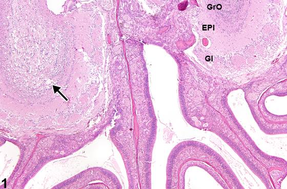
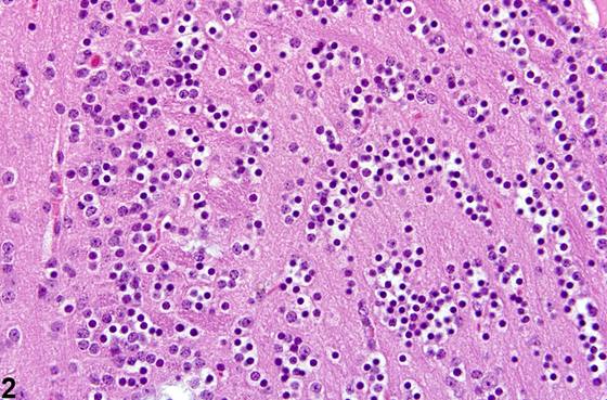
病理学家必须认识到与神经元损伤相关的各种形式的细微早期变化。这些类型的神经元变化可能在接受较低剂量受试化合物的动物中明显,或在较高剂量动物的脑内较不易受该药剂影响的核团中明显。神经组织损伤可能涉及一种或多种方式,例如血管损伤,其中液体在神经毡中积聚,造成该区域空泡化外观。直接神经元损伤可能涉及细胞膜以及离子和液体转移,在这种情况下,神经元核周体(perikaryon)中可能出现空泡。通常,损伤涉及代谢过程,特别是神经元能量产生的紊乱,在这种情况下发生细胞死亡,其特征为中央性染色质溶解(central chromatolysis)、神经元坏死,并最终溶解。Figure 1、Figure 2、Figure 3 在低倍和高倍放大下描绘了嗅球内颗粒细胞的变性。注意 Figure 1 中的变化在一个嗅球上更为严重(箭头),伴有神经元变性和丢失,以及整个嗅球结构的稀疏化。对侧嗅球通过薄骨板与受影响的对应部分隔开,嗅黏膜后部位于腹侧。对侧嗅球的结构在 Figure 1、Figure 2 中提供了良好的比较,具有正常的肾小球层、外丛状层和内颗粒细胞层。在毒理学研究中,密切观察嗅球的病变很重要,即使化合物不是通过吸入途径递送的。嗅球颗粒细胞的坏死和退行性变化在形态上与其他部位的颗粒神经元相似,例如小脑的颗粒细胞层。Figure 4、Figure 5 描绘了一种常被忽视的细微早期神经元损伤形式,称为“颗粒空泡变化(granulovacuolar change)”。核周体轻度肿胀,含有小空泡和颗粒状物质(箭头),提示膜损伤以及细胞质细胞器(如内质网和线粒体)的膨胀。在这些图像中,它伴有其他神经元损伤的辅助证据,例如中央性染色质溶解(黑色箭头)和真正的嗜碱性“慢性”神经细胞变化(白色箭头)。Cavanagh 等(1998)在一项关于真菌毒素对大鼠小脑影响的序贯研究中,提供了通过光镜和电镜记录的各种阶段的神经元和星形胶质细胞变性资料。
诊断与分级建议(NTP 共识)
当出现时,应诊断神经元变性,注明亚部位,并进行严重程度分级。在存在并发病变的情况下,通常诊断严重程度最高的病变。其他并发病变如果严重程度需要,可以单独诊断。
图谱
英文原文
展开 NTP 官方英文描述与建议
Comment:
It is important for the pathologist to be aware of the various forms of subtle early changes associated with neuronal injury. These types of neuronal changes may be evident in animals receiving lower doses of the test compound, or in nuclei of the brain less vulnerable to the agent from higher dosed animals. Injury to neural tissue may involve one or more modes, such as vascular injury in which fluid accumulates in the neuropil, creating a vacuolated appearance of the region. Direct neuronal injury may involve the membrane and ion and fluid transfer, in which case vacuoles may become evident in the neuronal perikaryon. Commonly, the injury involves metabolic processes, particularly disturbances to neuronal energy production, in which case cell death occurs characterized by central chromatolysis, neuronal necrosis, and ultimately lysis. Figure 1 , Figure 2 , and Figure 3 , at low and higher magnification, depict degeneration of internal granule cells of the olfactory bulb. Note that the change in Figure Figure 1 is worse on one olfactory bulb (arrow), with degeneration and loss of neurons, as well as rarification of the entire bulb structure. The contralateral olfactory bulb is divided from its affected counterpart by a thin osseous plate, and the posterior portion of the olfactory mucosa is present ventrally. The structure of the contralateral olfactory bulb provides a good comparison in Figure 1 and Figure 2 , having a normal glomerular layer, external plexiform layer, and internal granule cell layers. Close observation of the olfactory bulb for lesions is important in toxicologic studies, even if the compound is not delivered by the inhalation route. The necrotic and degenerative changes of granule cells of the olfactory bulb have a morphology similar to that seen in granule neurons in other sites, such as the granular cell layer of the cerebellum. Figure 4 and Figure 5 depict a commonly overlooked subtle and early form of neuronal injury known as “granulovacuolar change.” The perikaryon is slightly swollen and contains small vacuoles and granularity (arrowheads), suggesting membrane injury and distension of the cytoplasmic organelles such as the endoplasmic reticulum and mitochondria. In these images, it is accompanied by other ancillary evidence of neuronal injury, such as central chromatolysis (black arrows) and genuine basophilic “chronic” nerve cell change (white arrows). Various stages of neuronal and astrocytic degeneration as documented by light and electron microscopy are available in a sequenced study by Cavanagh et al. (1998) of the effects of a mycotoxin on the rat cerebellum.
Recommendation:
When present, neuron degeneration should be diagnosed, the subsite noted, and the severity graded. In the presence of concurrent lesions, lesions with the most severity are typically diagnosed. Other concurrent lesions may be diagnosed separately, if warranted by the severity.
来源与引用
原始条目:https://ntp.niehs.nih.gov/atlas/nnl/nervous-system/brain/Neuron-Degeneration
图谱总目录:NTP Nonneoplastic Lesion Atlas。 内容属美国联邦政府作品(公有领域),可引用;引用时请标注来源。
建议引用格式:NTP Nonneoplastic Lesion Atlas. Neuron Degeneration (brain). LabSmart 中文版:
https://www.labsmart.cn/ntp/brain-neuron-degeneration.html
中文图谱总页(可展开对照全部 595 条):https://www.labsmart.cn/ntp-atlas.html#brain-neuron-degeneration