神经元细胞丢失Neuron Cell Loss · 脑 · 神经系统 · NTP ntp-0391
诊断要点:在 NTP 研究中,任何检测到的神经元群体减少都应主观评定其严重程度,并诊断为神经元细胞丢失(Neuron, Cell loss)。
病变描述
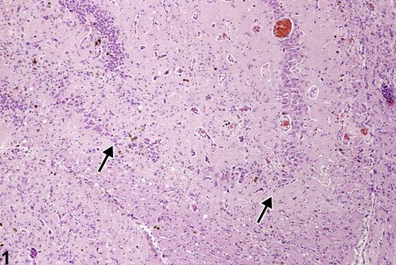
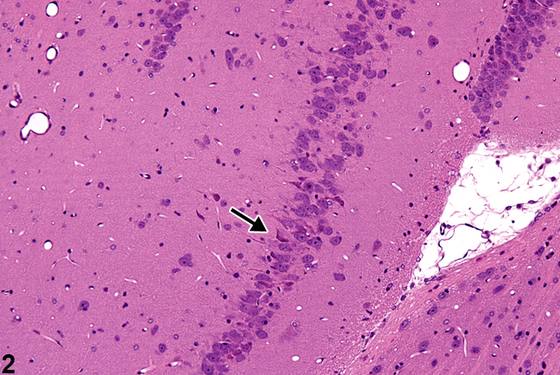
要检测神经元细胞丢失,必须了解大脑特定区域(海马、小脑)的正常神经解剖结构。普遍认为,常规主观光镜检查只能检测出细胞数量的显著减少。对更细微的丢失进行定量,需要采用胶质纤维酸性蛋白(glial fibrillary acidic protein)或 cresyl violet 特殊染色,或采用更精细的形态计量学和体视学技术。Figure 1 是海马 CA3 区神经元细胞丢失的一个示例。注意箭头之间锥体神经元的丢失,与邻近神经元丰富的区域形成对比。这是神经元坏死的晚期阶段。请将该图像与 Figure 2、Figure 3 中描绘的对照动物海马同一区域的图像进行比较。海马该部分的萎缩会干扰正常功能,尤其是学习、记忆和空间识别过程。必须将毒性损伤所致的神经元细胞丢失与区域性神经元发育不全(neuronal hypoplasia)及神经元萎缩(neuronal abiotrophy)相鉴别。神经元发育不全通常是自发性或诱发性发育异常,不伴有反应性胶质增生改变;而神经元萎缩是出生后发生的神经元丢失,呈进行性且与遗传相关,并可能有反应性改变。
诊断与分级建议(NTP 共识)
在 NTP 研究中,任何检测到的神经元群体减少都应主观评定其严重程度,并诊断为神经元细胞丢失(Neuron, Cell loss)。应注明受影响的脑亚部位并纳入描述。存在并发病变时,通常诊断严重程度最高的病变。其他并发病变若有充分依据,可根据其严重程度单独诊断。
图谱
英文原文
展开 NTP 官方英文描述与建议
Comment:
Knowledge of the normal neuroanatomic structure of specialized regions of the brain (hippocampus, cerebellum) is necessary to detect neuronal cell loss. It is generally accepted that routine subjective light microscopy detects only significant reductions in cell numbers. Quantitation of more subtle losses requires special staining with glial fibrillary acidic protein or cresyl violet or with more sophisticated techniques of morphometry and stereology. Figure 1 is an example of hippocampal neuronal cell loss in the CA3 region. Note the pyramidal neuronal loss between the arrows, in contrast to the adjacent neuron-rich region. This is a late stage of neuronal necrosis. Compare this image with those of Figure 2 and Figure 3 depicting the same region of hippocampus in a control animal. The atrophy of this portion of the hippocampus interferes with normal function, notably learning, memory, and spatial recognition processes. Neuronal cell loss due to toxic insult must be differentiated from regional neuronal hypoplasia and neuronal abiotrophy. Neuronal hypoplasia is usually a spontaneous or induced developmental abnormality unassociated with reactive gliotic changes while neuronal abiotrophy is postnatal neuronal loss that is progressive and genetically related and may have reactive responses.
Recommendation:
In NTP studies, any detected reduction in neuronal populations should be subjectively graded in severity and diagnosed as Neuron, Cell loss. Affected subsites of the brain should be noted and included in the narrative. In the presence of concurrent lesions, lesions with the most severity are typically diagnosed. Other concurrent lesions may be diagnosed separately, if warranted by the severity.
来源与引用
原始条目:https://ntp.niehs.nih.gov/atlas/nnl/nervous-system/brain/Neuron-CellLoss
图谱总目录:NTP Nonneoplastic Lesion Atlas。 内容属美国联邦政府作品(公有领域),可引用;引用时请标注来源。
建议引用格式:NTP Nonneoplastic Lesion Atlas. Neuron Cell Loss (brain). LabSmart 中文版:
https://www.labsmart.cn/ntp/brain-neuron-cell-loss.html
中文图谱总页(可展开对照全部 595 条):https://www.labsmart.cn/ntp-atlas.html#brain-neuron-cell-loss