神经元染色质溶解Neuron Chromatolysis · 脑 · 神经系统 · NTP ntp-0390
诊断要点:在 NTP 研究中,当存在染色质溶解时,应记录其解剖亚部位及严重程度分级。
病变描述
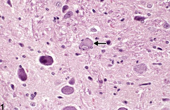
本图(Figure 1)显示的是神经元损伤中通常出现较早、往往为亚致死性的改变。它通常由毒物暴露及细胞代谢受干扰所致。就脊髓神经元而言,尤其可能继发于创伤性神经根受压或周围神经断裂。当神经元核周体中称为尼氏小体(Nissl substance)的粗面内质网及相关核糖体的正常聚集,作为对损伤的反应而分散时,即发生中央性染色质溶解(central chromatolysis,箭头)。它标志着在细胞损伤情况下神经元蛋白质合成的加速。与此同时,构成神经元细胞质骨架并支撑细胞核的细胞骨架网络受到不利影响。这种核悬浮的丧失导致细胞核失去其中心位置,变为偏位,紧邻细胞膜。在神经损伤恢复期间,尼氏小体可能重新聚集,但自中心开始,因此周边部细胞质仍保持透亮。这种改变称为“周边性染色质溶解(peripheral chromatolysis)”,被认为是早期神经元恢复的显微镜下指征。需要注意的是,脑内某些特别活跃的核团,如橄榄前核和下丘脑核团,其所含神经元正常情况下即具有明显的中央性染色质溶解,但无核偏位;因此,诊断这一改变时需谨慎。这就要求,无论中央性染色质溶解发生在何处,均应结合这些特定神经解剖部位的知识进行评估,并应与对照动物的相应部位进行比较,以避免对这一重要的神经元早期损伤特征发生误诊。
诊断与分级建议(NTP 共识)
在 NTP 研究中,当存在染色质溶解时,应记录其解剖亚部位及严重程度分级。
图谱
英文原文
展开 NTP 官方英文描述与建议
Comment:
This image ( Figure 1 ) displays the usually early, often sublethal, change that occurs in neuronal injury. It is commonly the result of toxin exposure and interference with cellular metabolism. In the case of spinal neurons, particularly, it may be secondary to traumatic radicular nerve crush or severance of the peripheral nerve. Central chromatolysis (arrow) occurs when the normal aggregations of rough endoplasmic reticulum and associated ribosomes, known as Nissl substance, in the neuronal perikaryon disperse as a response to injury. It signifies the acceleration of neuronal protein synthesis in the face of cellular injury. At the same time, the cytoskeletal network, which frames the neuronal cytosol and supports the nucleus, is negatively affected. This loss of nuclear suspension leads to the nucleus losing its central position, becoming eccentric, lying adjacent to the cell membrane. During recovery from the neural insult, the Nissl substance may reaggregate, but from the center, so that there remains cytoplasmic clarity peripherally. This change is referred to as “peripheral chromatolysis,” considered a microscopic indication of early neuronal recovery. It is important to note that some particularly active nuclei in the brain, such as the anterior olivary nucleus and hypothalamic nuclei, contain neurons that normally have apparent central chromatolysis, but not nuclear eccentricity; therefore, caution in the diagnosis of this change is required. This dictates that central chromatolysis, wherever it occurs, should be assessed with the knowledge of these particular neuroanatomic sites and should also be compared with equivalent sites in controls to avoid misdiagnosis of this important early neuronal feature of injury.
Recommendation:
When present, chromatolysis, its anatomic subsite location, and severity grading are noted in NTP studies.
来源与引用
原始条目:https://ntp.niehs.nih.gov/atlas/nnl/nervous-system/brain/Neuron-Chromatolysis
图谱总目录:NTP Nonneoplastic Lesion Atlas。 内容属美国联邦政府作品(公有领域),可引用;引用时请标注来源。
建议引用格式:NTP Nonneoplastic Lesion Atlas. Neuron Chromatolysis (brain). LabSmart 中文版:
https://www.labsmart.cn/ntp/brain-neuron-chromatolysis.html
中文图谱总页(可展开对照全部 595 条):https://www.labsmart.cn/ntp-atlas.html#brain-neuron-chromatolysis