上皮坏死Epithelium Necrosis · 鼻 · 呼吸系统 · NTP ntp-0123
诊断要点:上皮坏死只要存在就应诊断并分级。要诊断坏死,必须存在坏死细胞或其残余物。如果不存在坏死细胞,则应诊断溃疡或糜烂。
病变描述
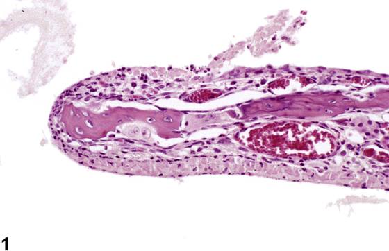
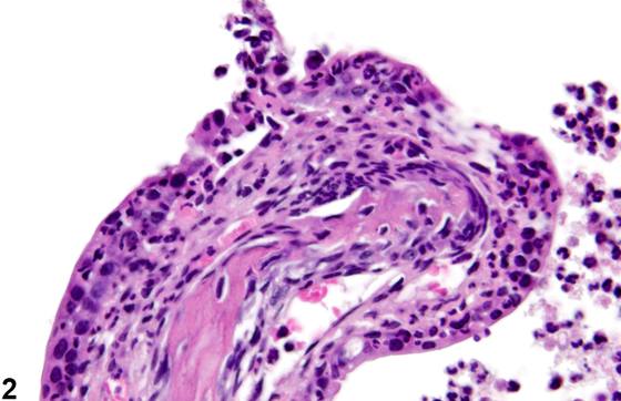
上皮坏死(epithelial necrosis,Figure 1、Figure 2、Figure 3、Figure 4、Figure 5、Figure 6)是吸入研究中常见的病变,尤其是在强刺激物作用下,但也可见于其他类型的研究。上皮坏死时可出现多种变化,包括细胞核固缩(pyknosis)或核碎裂(karyorrhexis)、核溶解(karyolysis)、细胞碎裂、胞质嗜酸性增强、细胞碎片的存在、细胞轮廓不清以及细胞脱落。任何上皮细胞或其他细胞类型均可受累,坏死可延伸至相关结构,如鼻腺、鼻泪管和鼻甲骨(Figure 6)。嗅上皮中感觉细胞的丢失常伴有固有层神经束的萎缩。
诊断与分级建议(NTP 共识)
上皮坏死只要存在就应诊断并分级。要诊断坏死,必须存在坏死细胞或其残余物。如果不存在坏死细胞,则应诊断溃疡或糜烂。诊断中应包含部位修饰词(例如呼吸上皮、移行上皮、鳞状上皮或嗅上皮、犁鼻器、泪管),以指示病变的位置。如果病变存在于一个以上的部位,则应针对其作为显著变化的每个部位分别诊断。坏死可与其他嗅部变化同时存在,当其作为独立、清晰明确的病变存在时应予以诊断。继发于其他病变(如炎症)的坏死性病变,除非严重程度足以支持,否则不应单独诊断,但应在病理学叙述中描述。相关病变,如炎性细胞浸润、炎症、鼻甲骨坏死或出血,除非严重程度足以支持,否则不应单独诊断。相关链接:鼻-上皮-变性;鼻-上皮-再生
图谱
英文原文
展开 NTP 官方英文描述与建议
Comment:
Epithelial necrosis ( Figure 1 , Figure 2 , Figure 3 , Figure 4 , Figure 5 , and Figure 6 ) a common lesion in inhalation studies, particularly with strong irritants, but may also be seen in other types of studies. A number of changes may manifest with epithelial necrosis, including pyknosis or karyorrhexis of nuclei, karyolysis, cell fragmentation, increased cytoplasmic eosinophilia, the presence of cell debris, indistinct cell outlines, and exfoliation of cells. Any of the epithelial or other cell types may be affected, and the necrosis may extend into associated structures such as the nasal glands, nasolacrimal ducts, and nasal turbinate bone ( Figure 6 ). Loss of sensory cells in the olfactory epithelium is frequently accompanied by atrophy of the nerve bundles in the lamina propria.
Recommendation:
Epithelial necrosis should be diagnosed and graded whenever present. The necrotic cells, or remnants thereof, must be present if necrosis is to be diagnosed. If necrotic cells are not present, then ulcer or erosion should be diagnosed. A site modifier (e.g., respiratory, transitional, squamous, or olfactory epithelium, vomeronasal organ, lacrimal duct) should be included in the diagnosis to indicate the location of the lesion. If the lesion is present in more than one location, it should be diagnosed separately for each location in which it is a prominent change. Necrosis may occur in the presence of other olfactory changes and should be diagnosed when present as a distinct, clearly defined entity. Necrotic lesions that are secondary to other lesions, such as inflammation, should not be diagnosed separately unless warranted by severity but should be described in the pathology narrative. Associated lesions, such as inflammatory cell infiltrate, inflammation, turbinate bone necrosis, or hemorrhage, should not be diagnosed separately unless warranted by severity. Related Links: Nose - Epithelium - Degeneration Nose - Epithelium - Regeneration
来源与引用
原始条目:https://ntp.niehs.nih.gov/atlas/nnl/respiratory-system/nose/Epithelium-Necrosis
图谱总目录:NTP Nonneoplastic Lesion Atlas。 内容属美国联邦政府作品(公有领域),可引用;引用时请标注来源。
建议引用格式:NTP Nonneoplastic Lesion Atlas. Epithelium Necrosis (nose). LabSmart 中文版:
https://www.labsmart.cn/ntp/nose-epithelium-necrosis.html
中文图谱总页(可展开对照全部 595 条):https://www.labsmart.cn/ntp-atlas.html#nose-epithelium-necrosis