斯滕森腺变性Steno's Glands Degeneration · 鼻 · 呼吸系统 · NTP ntp-0139
诊断要点:无论何时出现,Steno's glands 的变性、坏死和萎缩均应作出诊断并赋予严重程度分级。
病变描述
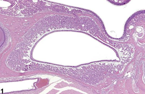
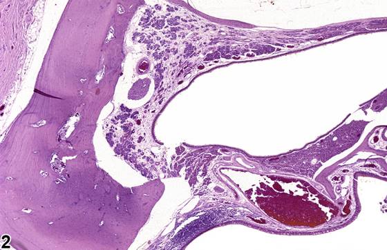
Steno's glands(斯坦诺腺),也称为侧鼻腺(lateral nasal glands),环绕上颌窦(maxillary sinuses)并延伸至外侧壁(Figure 1)。这些腺体的导管开口于鼻前庭(nasal vestibule)。在 mice 中,局灶性(focal) 萎缩可作为衰老性改变出现,但较为罕见。已知 Steno's glands 具有高代谢能力,可成为某些化学物的靶标。萎缩是细胞因坏死、凋亡或变性而丢失的最终结果,表现为腺体缺失或数量减少,或腺体皱缩(Figure 2、Figure 3、Figure 4)。通常,腺体导管仍然保留。腺体丢失或皱缩所留下的空间可能仅含有正常的细胞外基质(extracellular matrix),或者腺体可能被纤维化(fibrosis)所取代。Steno's glands 的坏死以出现核固缩(pyknotic nuclei)、胞质嗜酸性(cytoplasmic eosinophilia)、细胞轮廓不清(indistinct cell outlines),或细胞及细胞碎片脱落(exfoliation)为特征。变性被认为是细胞损伤的可逆形式,以细胞起泡(cell blebbing)、细胞排列紊乱(cellular disorientation)、细胞嗜酸性(cellular eosinophilia)、肿胀、细胞间接触丧失,或上皮内空泡化改变(vacuolar changes)为特征,并且也可能伴有炎症(inflammation)。变性和坏死可能伴有炎症。
诊断与分级建议(NTP 共识)
无论何时出现,Steno's glands 的变性、坏死和萎缩均应作出诊断并赋予严重程度分级。当 Steno's gland 组织量减少但几乎没有变性、坏死或炎症的证据时,应诊断为 Steno's glands 萎缩。如果存在上述变性或坏死的特征,则诊断应按此记录。如果同时存在炎症,不应单独诊断,除非其显著程度或严重程度足以支持单独诊断,但应在病理学描述(pathology narrative)中加以描述。
图谱
英文原文
展开 NTP 官方英文描述与建议
Comment:
Steno's glands, also called the lateral nasal glands, surround the maxillary sinuses and extend into the lateral wall ( Figure 1 ). The ducts of these glands empty into the nasal vestibule. Focal atrophy can be seen as an aging change in mice, but this is rare. Steno's glands are known to have high metabolic capacity and can be targeted by certain chemicals. Atrophy, the end product of cell loss due to necrosis, apoptosis, or degeneration, manifests as the absence or a decrease in number of glands, or shrunken glands ( Figure 2 , Figure 3 , and Figure 4 ). Often, the glandular ducts remain. The space left by the loss or shrinkage of the glands may simply contain the normal extracellular matrix, or the glands may be replaced by fibrosis. Necrosis of Steno's glands is characterized by the presence of pyknotic nuclei, cytoplasmic eosinophilia, indistinct cell outlines, or exfoliation of cells and cellular debris. Degeneration, which is considered to be reversible form of cell damage, is characterized by cell blebbing, cellular disorientation, cellular eosinophilia, swelling, loss of cell-to-cell contact, or vacuolar changes within the epithelium and also may be accompanied by inflammation. Degeneration and necrosis may be accompanied by inflammation.
Recommendation:
Whenever present, degeneration, necrosis, and atrophy of Steno's glands should be diagnosed and assigned a severity grade. Atrophy of Steno's glands should be diagnosed when the amount of Steno's gland tissue is decreased but there is little evidence of degeneration, necrosis, or inflammation. If the features of degeneration or necrosis described above are present, then the diagnosis should be recorded as such. If inflammation is present concurrently, it should not be diagnosed separately unless warranted by its prominence or severity, but should be described in the pathology narrative.
来源与引用
原始条目:https://ntp.niehs.nih.gov/atlas/nnl/respiratory-system/nose/Steno%27sGlands-Degeneration
图谱总目录:NTP Nonneoplastic Lesion Atlas。 内容属美国联邦政府作品(公有领域),可引用;引用时请标注来源。
建议引用格式:NTP Nonneoplastic Lesion Atlas. Steno's Glands Degeneration (nose). LabSmart 中文版:
https://www.labsmart.cn/ntp/nose-steno-s-glands-degeneration.html
中文图谱总页(可展开对照全部 595 条):https://www.labsmart.cn/ntp-atlas.html#nose-steno-s-glands-degeneration